31 May 2024
Dr. Ehab Shehata
Consultant Orthopedic Surgeon
Visit Profile
Book Appointment
Introduction:
This case study focus on effective, successful management and treatment of challenging difficult complex left tibial pilon in 32 – years-male, at the Department of Orthopedic surgery, International Modern hospital. It showcase the hospital commitment to the excellence of patient care and the utilization of advanced Minimal invasive Orthopedic Procedures and techniques.
Patient Background:
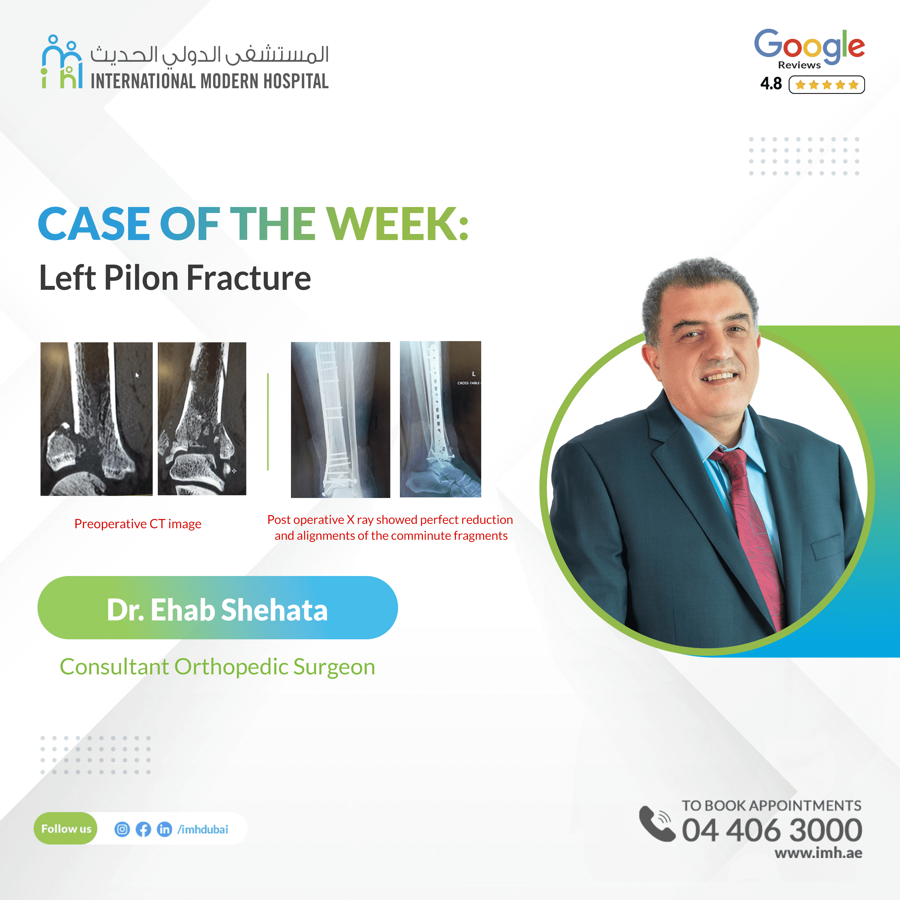
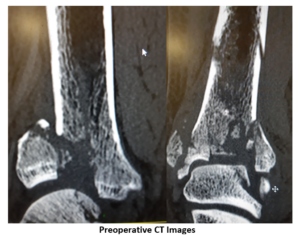
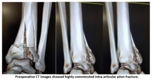
The patient come to ER after RTA with massive trauma with left ankle sever edema and inability to standing or walking. X ray then Ct was done, showed complex intra articular comminuted displaced Pilon fracture with articular surface depressed and fragmented left lower tibia and fibula complex intra articular fracture since three days, patient was in another hospital and shifting to our hospital, for CT and preparation for ORIF
Background:
These fractures account for approximately 1% to 10% of the lower leg or tibial fractures and are often associated with severe bone comminution and soft tissue compromise. Pilon fractures may also involve metaphyseal extension and can have associated fibular fractures.
The distal tibia has a quadrilateral cross-sectional shape and together with the fibula, ligaments, and capsule, forms the ankle mortise. This topography is designed to maximize the articular surface area with the dome of the talus and minimize the stress on the ankle joint.
The tibia and fibula are held together by the interosseous membrane, anterior inferior and posterior inferior tibiofibular ligaments. The vascular supply of the tibial plafond derives from branches of the anterior tibial, posterior tibial, and peroneal arteries
Mechanism of Fracture:
Pilon fractures most often result from high-energy mechanisms, such as motor vehicle accidents and falls from a height , The impact from an axial compression mechanism drives the articular surface proximally into the metaphysis, with associated metaphyseal comminution.
The anterior or Chaput fragment is attached to the anterior inferior tibiofibular ligament (AITFL); the posterior or Volkmann fragment is attached to the posterior inferior tibiofibular ligament (PITFL); the medial malleolar fragment is attached to the deltoid ligament. Fragments that maintain their normal alignment or fractures without this typical Y-shaped pattern may be indicative of ligament rupture the Pilon fracture is well-known as one of complex and challenging fracture for operative treatment for experiencing Orthopedic surgeons because of very difficult to achieve Anatomical Reduction, Associated soft tissue injury, Usually required multiple surgeries to achieve proper heeling
Operative Technique:
The first step requires the restoration of fibular length to re-establish the lateral column, which aids in the reduction of the tibial plafond. The second step calls for anatomic restoration of the articular surface of the distal tibia, which often resembles a jigsaw puzzle given the severe comminution. Lastly, a buttress plate is placed on the distal aspect of the tibia.
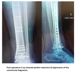
Minimal invasive techniques was used in this case with distal medial approach and achieved good articular alignment then primary fixation by KW the subcutaneous long Special MEPO Plate fixation technique was done, and final stable rigid bone-implant construct was achieved.
Patient tolerated surgical procedure (three hours times), , and next day patient was discharged in below knee splint.
Conclusion:
Minimal invasive surgery consider as advanced techniques in orthopedic surgery and MEPO fixation in those comminuted changing fracture considering as one of the best results and minimal complications rate and need high experience surgeon to perform it.


