17 May 2024
Dr. Ehab Shehata
Consultant Orthopedic Surgeon
Visit Profile
Book Appointment
Introduction:
This case study focus on effective, successful management and treatment of challenging difficult complex Right elbow fracture dislocation with sever displaced intra articular fracture in 41 years male, at the Department of Orthopedic surgery, International Modern hospital. It showcase the hospital commitment to the excellence of patient care and the utilization of advanced Minimal invasive Orthopedic Procedures and techniques.
Patient Background:
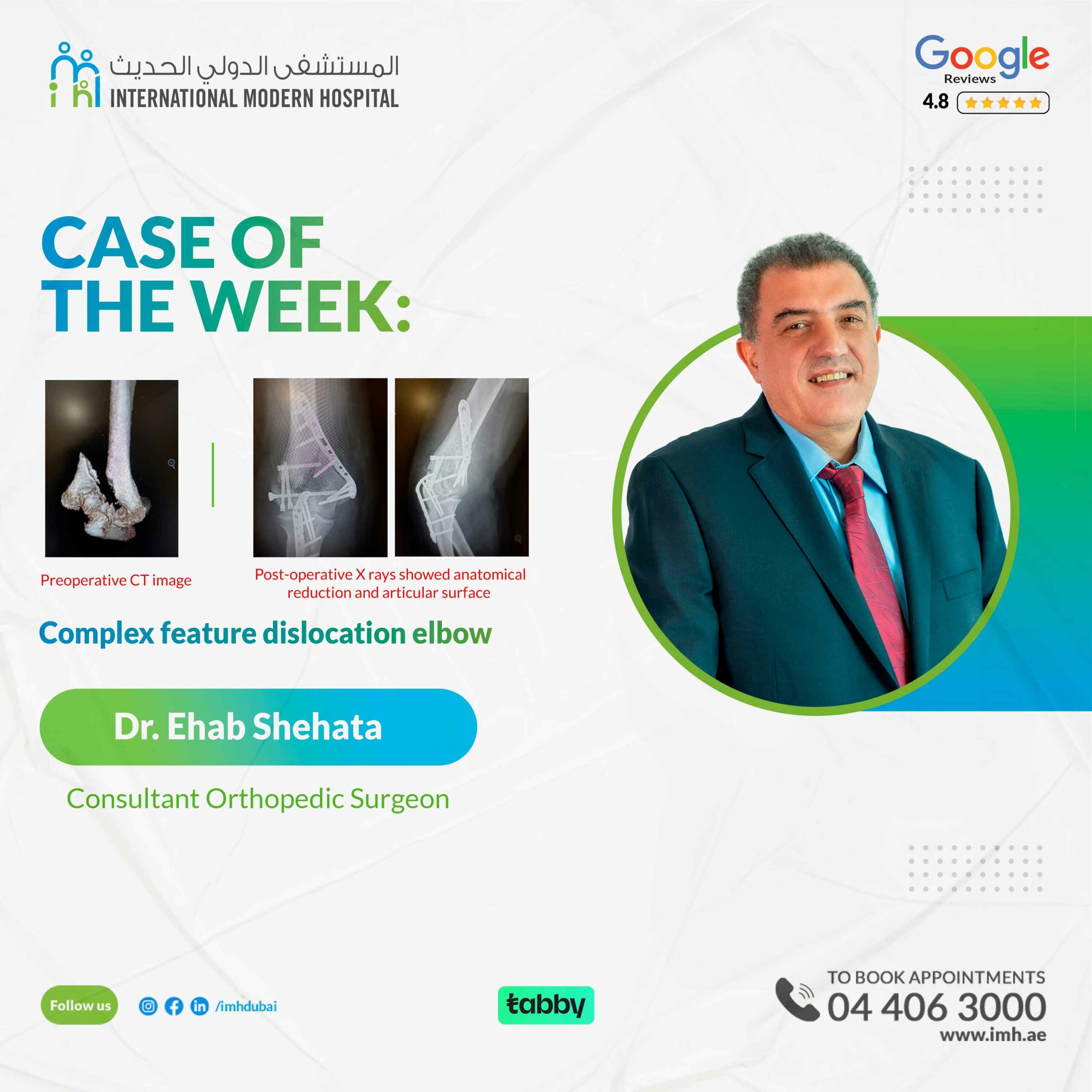
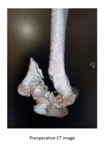
The patient come to ER after falling down direct over right elbow joint, with massive trauma with Right elbow sever pain, swelling and weak radial pulsation and inability to move elbow and hand. First aid splint and urgent X ray and CT was done, showed complex intra articular comminuted displaced intra- articular distal humeral fracture with sever ligament tears and disrupted articular surfaces involving capitllum and Trochlea
Background:
Elbow fracture dislocations are complex injuries that can provide a challenge for experienced surgeons. Current classifications fail to provide a comprehensive system that encompasses all of the elements and patterns seen in elbow fracture dislocations.
Elbow stability is a function of osseous and ligamentous constraint and musculotendinous control. The contribution of each is not equal across the whole elbow. On the medial side, the olecranon and coronoid processes enclose the medial trochlea, producing a high degree of concavity compression or constraint, which means that the ligaments and musculotendinous units on the medial side are of lesser importance. Laterally the radial head is relatively incongruent with a greater arc of curvature than the capitellum, the static and dynamic soft tissue restraints of ligaments and tendons are of much greater importance, and detachment of these soft tissue structures are more likely to result in symptomatic instability.
Despite advances in radiographic imaging, surgical instrumentation, and our understanding of the importance of soft tissue handling, Those fractures remain challenging to treat. Clinical outcomes correlate with severity of the fracture pattern and the quality of reduction.
The Neuro- Vascular injuries was more common in those trauma included Ulnar, radial and median nerve with brachial artery with high incidence of immediate or later complications affecting the movements of the hand and elbow, with high incidence of long term/ persistence stiffness and numbness.
Complications after surgical fixation include wound slough or dehiscence, infection, malunion, nonunion, joint stiffness, and post-traumatic arthritis remain major challenges.
Mechanism of Fracture:
Elbow comminuted fractures most often result from high-energy mechanisms. The impact from an axial compression mechanism drives the articular surface proximally into the metaphysis, with associated metaphyseal comminution the elbow fracture dislocation is well-known as one of complex and challenging fracture for operative treatment for experiencing Orthopedic surgeons because of very difficult to achieve Anatomical Reduction, Associated soft tissue injury, Usually required multiple surgeries to achieve proper heeling and long term follow up and physiotherapy to achieve proper ROM and factional activity.
Operative Technique:
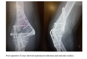
The first step requires , trans olecranon osteotomy approach with identification and anterior transfer of the ulnar nerve, to expose the articular distal humeral fragments.. the Assembly the fragments then checked by C-arm, then primary fixation by Kw then final by inter-fragmentary screws restoring the articular surface in perfect alignment then the lateral Colman with special locked plate and lastly the medial Colman with cannulated screws, to get finally stable rigid bone implant construct. Allowed ROM 0-0-110 intra operatively.
Patient tolerated surgical procedure (three hours times), , and next day patient was discharged in above elbow splint, and weekly follow up, after two weeks patient removed the splint and wearing special Elbow ROM brace with allowed ROM 90 flexion –extension range to reached to 100 Range of motion with in 4 weeks post operative and started early physiotherapy.
Conclusion:
Anatomical reduction and stable rigid internal fixation followed by early ROM considered as best techniques in orthopedic surgery in those comminuted challenging fracture considering as one of the best results and minimal complications rate and need high experience surgeon to perform it.


